
5th International Andrology Symposium
5th International Andrology Symposium
Antoine H.F.M. Peters (Basel)
Friedrich Miescher Institute for Biomedical Research, 4056 Basel, Switzerland; Faculty of Sciences, University of Basel, 4056 Basel, Switzerland
Correspondence; Adjunct Professor of Epigenetics, University of Basel, Basel Switzerland
Male germ cell development unfolds through a sequence of lineage transitions—primordial germ cell (PGC) specification and reprogramming, prospermatogonial (gonocyte) quiescence and conversion to spermatogonial stem cells (SSCs), amplification/differentiation of spermatogonia, and meiosis followed by spermiogenesis—that are choreographed in part by epigenetic mechanisms. DNA methylation is globally erased in PGCs and later re-established de novo in the fetal/neonatal male germ line; histone post-translational modifications (PTMs) and Polycomb-group (PcG) repressive systems create chromatin environments that both permit and restrict gene expression programs; the piRNA pathway safeguards genomic integrity by directing transposable-element silencing. Using lessons from our laboratory’s work on chromatin-based memory in gametes and early embryos, and integrating recent insights from male germ cell epigenomics, I discuss: (i) how PcG complexes (PRC1/PRC2) and their readers/writers organize developmental competence in spermatogonia; (ii) how CpG-island (CGI) methylation is protected during oogenesis by KDM2A/KDM2B to prevent aberrant maternal hypermethylation that escapes embryonic reprogramming (a paradigm for how epigenetic promoter chromatin states can be inherited); (iii) the emerging view that H3K27me3/H2AK119ub landscapes are dynamically regulated in undifferentiated spermatogonia and may be perturbed by intrinsic or environmental stressors resulting in cryptorchidism; and (iv) opportunities to apply single-cell multi-omics to cryptorchid testes to resolve whether a failed developmental transition from gonocytes to Adark SSC reflects the acquisition of intrinsic epigenetic roadblocks. I conclude by outlining a framework in which developmental “flexible repression” by Polycomb must be maintained to keep germline genes poised, while inappropriate switching to DNA-methylated locked repression at key promoters may derail fate transitions with clinical consequences for fertility.
Key words: Epigenetic, germ cell Polycomb group
Le développement des cellules germinales mâles progresse par une succession de transitions lignagères—spécification et reprogrammation des cellules germinales primordiales (PGCs), quiescence des prospérmatogonies (gonocytes) et conversion en cellules souches spermatogoniales (SSCs), amplification et différenciation des spermatogonies, puis méiose et spermiogenèse—régulées en partie par des mécanismes épigénétiques. La méthylation de l’ADN est globalement effacée dans les PGCs puis rétablie de novo dans la lignée germinale fœtale/néonatale ; les modifications post-traductionnelles des histones et les systèmes répressifs Polycomb façonnent des états chromatiniens permissifs ou restrictifs ; le système piRNA protège l’intégrité génomique par la répression des éléments transposables. À partir de travaux de laboratoire sur la mémoire chromatinienne dans les gamètes et l’embryon précoce, ainsi que des avancées en épigénomique germinale masculine, nous examinons : (i) le rôle des complexes Polycomb (PRC1/PRC2) dans l’établissement de la compétence développementale des spermatogonies ; (ii) la protection de la méthylation des îlots CpG durant l’ovogenèse par KDM2A/B, évitant une hyperméthylation maternelle échappant à la reprogrammation embryonnaire ; (iii) la régulation dynamique des marques H3K27me3/H2AK119ub dans les spermatogonies indifférenciées et leur possible perturbation par des stress intrinsèques ou environnementaux, notamment dans la cryptorchidie ; et (iv) l’intérêt des approches multi-omiques unicellulaires pour déterminer si l’échec de conversion gonocyte → SSC Adark reflète des blocages épigénétiques intrinsèques.
Nous proposons un modèle où la “répression flexible” assurée par Polycomb doit être maintenue pour garder les gènes germinatifs en état de veille, tandis qu’une transition inappropriée vers une répression verrouillée par méthylation de l’ADN peut compromettre les trajectoires cellulaires, avec des conséquences cliniques pour la fertilité.
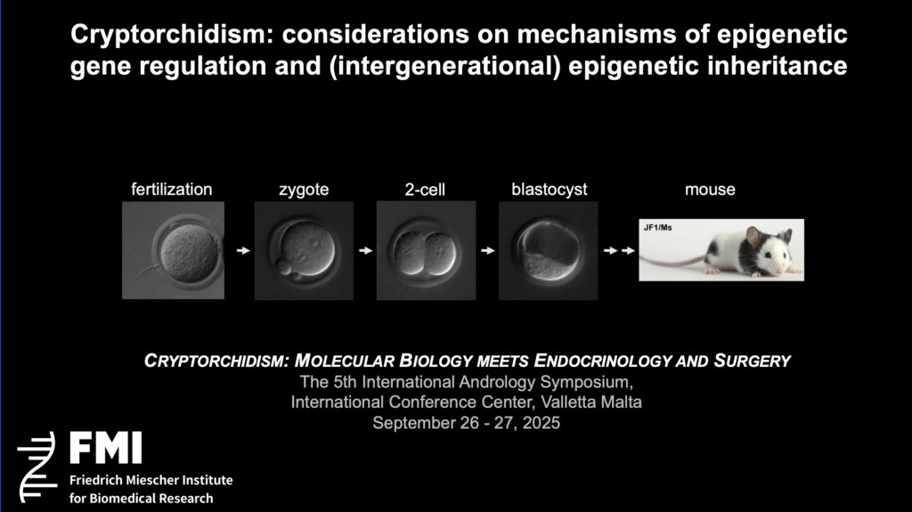
We examined how transcription factors and chromatin pathways establish, memorize, and transmit gene-regulatory states across cell divisions and between organismal generations, with a focus on mammalian gametes and preimplantation embryos [1]. We uncovered how histone modifiers gate de novo DNA methylation and how Polycomb-mediated repression prevents an inappropriate conversion to hardwired DNA methylation—are highly relevant to male germ cells. In this proceedings contribution I connect these mechanistic principles to spermatogenic development, highlight new evidence on PcG regulation of undifferentiated spermatogonia, and propose how these insights may illuminate cryptorchidism-associated defects in the gonocyte-to-SSC transition described clinically and transcriptionally by others [2,3].
Chromatin encodes two broad, functionally distinct states of gene silencing.
A central lesson from our lab is that choices made in the female germ line can be transmitted to the embryo. In mouse oocytes, the demethylases KDM2A/KDM2B restrain H3K36me2 at CGIs, preventing DNMT3A from methylating promoters. If KDM2A/KDM2B proteins are absent, aberrant hypermethylation accumulates at CGI promoters in growing oocytes, persists into four-cell embryos, and represses the maternal allele of key developmental genes. Strikingly, removal of DNMT3A in the oocyte rescues the preimplantation lethality of Kdm2a/Kdm2b deficient embryos, proving causality [9]. Although discovered in oocytes, the principle—that the histone mark balance at promoters gates access of DNMT3A, determining whether repression stays Polycomb-flexible or becomes DNA-methylation-locked—applies to spermatogonia as well [11].
PGCs undergo global DNA demethylation (active and passive) during migration and gonadal colonization, removing parental methylation genome-wide including at CGIs, imprinting control regions and many endogenous repetitive viral elements. In the male germline, prospermatogonia acquire high levels of de novo methylation during late fetal/neonatal stages under the control of de novo DNA methyltransferases DNMT3A, DNMT3C and DNMT3L. This re-methylation licenses future spermatogenesis and transposon restraint, in part in cooperation with the piRNA pathway (MILI, MIWI2 and cofactors) [12–15].
Undifferentiated spermatogonia (including SSCs) exhibit PcG-enriched chromatin: PRC1/PRC2 partition developmental regulators into a repressed-but-poised compartment characterized by H3K27me3 and H2AK119ub. During commitment to differentiation, global H3K27me3 levels decrease and specific PcG domains are remodeled, allowing lineage genes to be activated with proper timing [4–7]. In mouse, PRC1 constrains premature activation of germline genes and coordinates their timely expression during spermatogenesis; PRC1 loss disrupts this choreography [4,5]. In adult mice, PRC1 shields undifferentiated spermatogonia from stochastic differentiation, helps maintain slow cycling, and directs orderly commitment; PRC2-H3K27me3 emerges as an epigenetic hallmark of this population [4-6].
Entering meiosis entails broad transcriptional reprogramming with stage-specific histone marks (e.g., H3K4me3, H3K9me3, H3K27me3 dynamics) and recruitment of germline TFs. Post-meiotically, massive histone eviction and protamine incorporation fully reprograms the paternal epigenome. Nonetheless, few nucleosomes are retained in sperm, the function of which remains to be identified [16-20]. PcG factors (e.g., SCML2) and modulators such as EZHIP can tune PRC2 activity in gonads, with consequences for H3K27me3 abundance during spermatogenesis [21].
In mESCs and in vivo, H2AK119ub (PRC1) often shows rapid restoration after DNA replication, whereas H3K27me3 (PRC2) accumulates more slowly, suggesting that PRC1 can prime domains for PRC2 spreading [22]. In male germ cells, convergent evidence indicates that PRC1 establishes and maintains repressive compartments in undifferentiated spermatogonia, preventing inappropriate transcription and preserving stemness; during differentiation, selected PcG domains are dismantled so germline programs can proceed [4–6,11].
If PcG repression is weakened, developmental and pro-apoptotic genes become ectopically activated [7]. If repression is inappropriately hardened by promoter DNA methylation, a different pathology arises—failure to activate required genes when differentiation cues arrive [10]. Either imbalance may also prevent the gonocyte to Adark SSC transition observed in cryptorchid testes, where failure to establish the Adark reserve population presages subfertility [2]. A crucial inference is that maintaining Polycomb-facilitated flexibility, not converting promoters into DNA-methylated hard-locks, might be critical in this window.
A surgically induced cryptorchid model has recently been established in adult mice to profile the epigenetic landscape of undifferentiated spermatogonia. The key observation is a local (not global) loss of H3K27me3 and H3K9me3 at PcG-regulated genes, accompanied by upregulation of developmental and pro-apoptotic pathways. Cryptorchid spermatogonia show increased expression of H3K27 demethylases KDM6A/KDM6B, and elevated temperature directly induces Kdm6a/Kdm6b in germline stem cell culture [7]. These data dovetail with earlier work linking abnormal H3K27me3 to impaired SSC maintenance and differentiation [4]. They point to a plausible mechanism whereby aberrant scrotal heat exposure—central to cryptorchidism—erodes PcG-mediated flexible repression, de-repressing stress-response and developmental genes, precipitating either apoptosis or mis-timed differentiation. Whether a similar chromatin response is observed in developmentally cryptorchid juvenile mice, and whether subsets of promoters may subsequently convert to DNA-methylated locked states remains to be tested in longitudinal models.
Our 2025 studies establish that the KDM2A/KDM2B histone demethylases, binding to unmethylated CpG islands, remove H3K36me2 and thereby limit DNMT3A access to promoter CGIs in growing oocytes. In their absence, widespread CGI hypermethylation arises which is inherited to four-cell embryos, where it leads to allele-specifically transcriptional repression from the maternal genome. Deleting Dnmt3a in oocytes rescues embryonic lethality, proving that aberrant DNA methylation is the causal lesion [10]. Mechanistically, we further uncover a promoter “grammar” in which the antagonism between H3K4 methylation (protective) and H3K36 methylation (permissive for DNMT3A) and local sequence features determines whether a promoter flips from Polycomb to DNA methylation when KDM2A/KDM2B protection is removed [10].
While male prospermatogonia do undergo physiologic de novo methylation, the hypothesis is that many stem/progenitor promoters in undifferentiated spermatogonia must remain unmethylated and Polycomb-repressed to preserve flexibility [6, 11]. Inappropriate promoter methylation—due to chronic stress, imbalance of chromatin modifying enzymes (e.g., altered KDM2 family function, excessive H3K36 signals), or mis-coupling of PRC1/PRC2—could permanently lock out essential fate genes [11]. Single-cell bisulfite DNA methylation and chromatin modification profiling via CUT&TAG/CUT&RUN studies in human cryptorchid testes could test whether high-risk infants show aberrant CGI hypermethylation (locked repression) at Adark-associated gene sets.
Retrotransposons (LINE-1, IAP, etc.) are silenced in male germ cells via piRNA-directed DNA methylation and repressive histone marks. Failure of the MILI/MIWI2 axis compromises genome integrity and induces spermatogenic arrest. Notably, Polycomb and piRNA systems intersect functionally: PcG repression can help buffer developmental genes while piRNA machinery targets repeats, collectively allowing germ cells to proceed through meiosis without genotoxic stress [6,11-14]. Temperature stress may also perturb piRNA biogenesis; integrating small RNA profiling with PcG mapping in cryptorchid models should reveal whether TE de-repression contributes to the observed SSC attrition.
Clinical and histological studies indicate that cryptorchidism with high risk for adult infertility is characterized by the absence of Adark spermatogonia—the reserve SSC pool in infant boys. Expression profiling of infant testes stratified by Adark status shows widespread low expression of genes implicated in SSC identity and differentiation, consistent with a blocked transition from gonocyte (prospermatogonia) to SSC [2,3]. Re-interpreting such signatures through an epigenomic lens suggests two, not mutually exclusive, failure modes:
These models make distinct predictions. While the PcG erosion model yields loss of H3K27me3 (without DNA methylation change) at specific loci, the lock model yields new promoter CGI methylation. Single-cell multi-omics on pediatric cryptorchid testes can discriminate them.
The testis is a mosaic of germ and somatic cells. Bulk profiling can be misleading. Single-cell atlases in the human testis have already delineated SSC sub-states and somatic niches. Extending them with single-cell CUT&TAG (H3K27me3/H2AK119ub), single-cell methylome, and small-RNA sequencing in cryptorchid and control testes (correcting for age, temperature exposure, and surgery timing) will identify cell-intrinsic chromatin lesions in germ cells versus extrinsic niche signals (Sertoli, Leydig, peritubular myoid). A prospective study that pairs longitudinal sampling with orchiopexy timing could test whether early surgery prevents PcG erosion and reduces promoter locking at SSC-critical genes.
Our oocyte studies show that “flexible” Polycomb repression can be converted to “rigid” DNA-methylation repression at promoters if KDM2A/KDM2B-mediated protection is lost; this locked repression is heritable into the embryo and causally impairs development [10]. In male germ cells, we posit that the same decision logic applies to promoters of SSC competence and differentiation genes. Meanwhile, PRC1/PRC2 keep undifferentiated spermatogonia poised; when disrupted (by genetics or environment), timing and survival are perturbed [4–7]. Finally, modulators such as EZHIP can dampen PRC2 activity in the gonads, reminding us that PcG dosage is tightly tuned in the germ line [21]. Bringing these strands together, a unifying epigenetic model of male germ cell development emerges: Polycomb builds the poised male germ line landscape; KDM2A/KDM2B-guarded CGIs keep promoters unmethylated; DNMT3A writes stable locks where appropriate (endogenous repetitive elements, imprints but not at SSC-critical promoters); piRNA system patrols repeats; and temperature or inflammation stress can upset the balance. [22].
This proceedings’ contribution synthesizes published primary data and reviews, including mechanistic experiments from our laboratory and others. Where appropriate, I refer to mouse models (oocyte Kdm2a/Kdm2b, Dnmt3a; spermatogonial PcG perturbations; cryptorchid surgery) and to human single-cell atlases and clinical observations. No new datasets are presented here; rather, I propose testable hypotheses and study designs (single-cell multi-omics of cryptorchid testes) grounded in the cited literature.
Male germ cell development depends on a choreography of chromatin states: Polycomb maintains a poised genome in undifferentiated spermatogonia; piRNA and DNA methylation secure the genome against transposons; and promoter CGI protection prevents conversion to irreversible repression. The recent demonstration that promoter choice in the oocyte can be memorized into the embryo provides a strong precedent for similar modes of regulation in the male germ line. In cryptorchidism, temperature-induced PcG erosion and potential promoter locking offer mechanistic explanations for the failure of the gonocyte to Adark transition. The immediate priority is to map PcG marks, promoter methylation, and small RNAs at single-cell resolution in infant testes at risk. The medium-term goal is to determine whether restoring the poised state can preserve future fertility.
a) Ethics Approval and Consent to Participate Investigations were carried out in accordance 326 with the Declaration of Helsinki of 1975, revised in 2008.
b) Consent for publication Not applicable
c) Availability of data and supporting material Not applicable
d) Competing interests Author/s declare that they have no competing interests
e) Funding none
I thank Y. Kawamura, E. Ozonov, P. Papasaikas and all past and present members of my group for their contributions; H. Koseki for essential genetic mouse models; and our collaborators at FMI Basel for continuous support.

Lorem ipsum dolor sit amet consectetur. Id mollis nulla maecenas at vestibulum blandit consectetur. Vulputate libero turpis diam eu rhoncus arcu. Donec at imperdiet viverra ut eu sagittis nunc volutpat. Sem nisi turpis venenatis non sed adipiscing donec dignissim.